Daily medical practice is facing a new era of development with the emergence of Artificial Intelligence (AI). This digital revolution in medicine has the potential to transform the manner in which care is provided by becoming an effective tool to help clinicians in daily practice. Moreover, the impetus within AI advances is not only exclusive to improve the performance and the accuracy of clinical tests, but also to reduce costs.
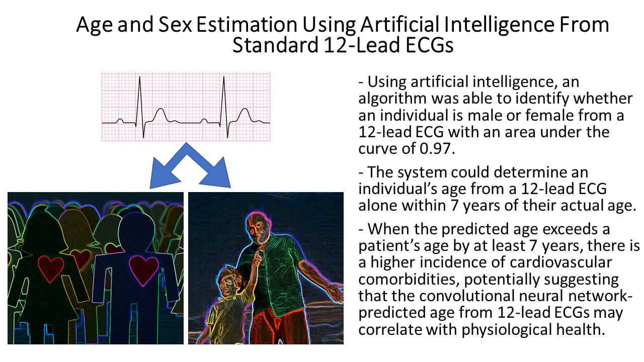
 Recently, a Mayo Clinic team reported the added value of AI in identifying a number of physiological variables (1). As already known, AI techniques may correlate features of an ECG with specific phenotypic findings (2,3). In fact, the aim of their study was to use AI to determine age and sex reported by patients from a standard 12-lead ECG, and to determine whether discrepancies between ECG age and chronological age might be a marker of physiological health. A convolutional neural network (CNN) was trained, through a process called deep learning, using 10-second samples of 12-lead ECG signals from 499,727 patients to predict sex and age (figure). The network was then tested on a data set comprising of 275,056 patients. Here after, 100 randomly selected patients with multiple ECGs over a minimum period of 2 decades were used to assess the accuracy of the CNN age estimation. Finally, the incidence of pre-existing comorbidities was assessed. The results of this study showed encouraging data that a CNN could be trained to predict with reliable accuracy the sex and age of a patient Furthermore, low ejection fraction, hypertension and coronary disease have been identified with a higher incidence among patients with a CNN-predicted age that exceeded chronological age by over 7 years
As inherent for most single centre datasets, some limitations in the Attia paper are evident. For example, the incidence of cardiovascular diseases in the population used to develop the algorithm has not been reported. Furthermore categorisation in terms of race and age groups has not been described leading to potential disparities in data representation. Additionally the authors randomly selected 100 patients with longitudinal data (patients with multiple ECGs recorded over a minimum of 20 years) with the objective to determine the gap between CNN-predicted age and chronological age and to evaluate if pre-existing comorbidities were more frequent among patients whose CNN-predicted age exceeded the chronological age. All these issues raise the limitation of a possible selection bias, as patients undergoing multiple ECGs plausibly encounter more underlying comorbidities. These questions have been addressed by the authors, but remain unanswered until now.
Moreover the opaque nature of the “Black Box” within AI derived outcomes remains contentious. In the Attia paper, the algorithm cannot explain which characteristics of the ECG morphology led to a specific result. Any attempt to interpret Attia’s black box is in essence of inconsequential significance when one assumes that the neural network is applied for screening of a cardiovascular condition. One must also consider that neural networks are contingent on data quality and quantity which in itself amplifies the complexity of these models.
Another frequently cited argument is the potential for disparity of the ECG recordings, particularly in the context of electrode positioning which are prone to human errors and some cases non-uniform electrode placement (4). This argument has been recently extensively detailed by Van Dam et al (5) in which the benefit of using a three-dimensional camera for electrode placement has been proposed to help ensure a standard ECG output.
The current debate leads one to deliberate that the impact of this particular algorithm on our daily clinical practice intuitively warrants further investigation. However it could be postulated that ECG analysis by AI could be used in everyday practice by general practitioners as a screening tool for indiscriminate latent cardiovascular diseases in the general population (6). Furthermore it may be of interest in remote areas, where specialized medicine is less easily accessible. Hence one could reasonably qualify the promise to improve risk stratification, speed up individualized medical care and thus positively impact long-term outcome. Notwithstanding , the added value of this AI technique on a specific and thus well-defined cardiovascular population of patients remains questionable, given the necessity for a meticulousness and prompt diagnosis of underlying disease.
To conclude, the use of AI in medicine continues to exhibits promise. However, the exact connotation and significance of AI in current practice warrants further study and clarification.
Conflict of Interest
Nothing to declare


 Our mission: To reduce the burden of cardiovascular disease.