Background
HCM is a common genetic cardiovascular disease with an overall prevalence estimated between 0.05-0.2% of the population. (1) HCM is a disease state characterised by unexplained, marked and asymmetric left ventricular (LV) hypertrophy associated with non-dilated ventricular chambers in the absence of another cardiac or systemic disease capable of producing the magnitude of hypertrophy evident in a given patient. (2) Clinically, HCM is usually recognised by a maximal LV wall thickness >15 mm. However, it should be underscored that, in principle, any degree of wall thickness is compatible with the presence of the HCM genetic substrate.
1 - Approach
In order to establish the diagnosis of HCM, a systematic echocardiography approach is necessary. The echocardiographic examination should include:
- Confirming LV hypertrophy
- Assessment of LVOT obstruction
- Systolic anterior motion - SAM
- Assessment of systolic and diastolic LV function and left atrial size
Additional novel echocardiographic modalities make for better understanding of subclinical LV damage. Transthoracic echocardiography is recommended as a component of the screening algorithm for family members of patients with HCM.
2 - Confirming the presence/absence of left ventricular hypertrophy
Hypertrophy preferentially involves the interventricular septum in the basal LV segments, but often extends into the lateral wall, posterior septum and LV apex. Although HCM is typically characterised by asymmetric septal hypertrophy (ASH), almost any myocardial segment may be involved.
The following two-dimensional (2D) echocardiographic criteria are used to aid diagnosis:
- Unexplained maximal wall thickness >15 mm in any myocardial segment, or
- Septal/posterior wall thickness ratio >1.3 in normotensive patients, or
- Septal/posterior wall thickness ratio >1.5 in hypertensive patients.
Nevertheless, genotype positive adults (including those who die suddenly) may have normal or near normal wall thickness. Assessing the extent and severity of hypertrophy must include the measurement of maximal wall thickness in all LV segments from base to apex, ensuring that the wall thickness is recorded at mitral, mid-LV and apical levels. It is important to recognise, however, that asymmetric left ventricular hypertrophy (LVH) in itself is not pathognomonic of HCM, but may be encountered in a variety of congenital or acquired conditions, including right ventricular hypertension, systemic hypertension, aortic stenosis, septal sarcomas, Fabry's disease, Freidreich's ataxia, mucopolysaccharide or glycogen storage disorders and amyloidosis.
Associated abnormalities of left ventricular outflow tract: the pathophysiology of HCM is complex and consists of multiple interrelated abnormalities, including LVOT obstruction, diastolic dysfunction, mitral regurgitation, myocardial ischemia, and arrhythmias. (4) It is clinically important to distinguish between the obstructive and nonobstructive forms of HCM because management strategies are largely dependent on the presence or absence of symptoms caused by obstruction.
For HCM, it is the peak instantaneous LV outflow gradient, rather than the mean gradient, that influences treatment decisions. Up to one-third of patients with HCM will have obstruction under basal (resting) conditions (defined as gradients ≥30 mm Hg). Another one-third will have labile, physiologically provoked gradients (<30 mm Hg at rest and ≥30 mm Hg with physiologic provocation). (5) The final one-third will have the nonobstructive form of HCM (gradients <30 mm Hg at rest and with provocation). Marked gradients ≥50 mm Hg, either at rest or with provocation, represent the conventional threshold for surgical or percutaneous intervention if symptoms cannot be controlled with medications.
The presence of SAM is documented using M-mode echocardiography and is characterised by mid-systolic notching of the aortic valve and contact of the anterior mitral valve leaflet/chordae with the septum. In obstructive HCM, IVS is much wider, with smaller LV cavity and greater incidence of mitral insufficiency, generally directed towards the posterior wall of the left atrium, and a larger tendency to calcify the mitral annulus. Obstruction to LV outflow is dynamic, varying with loading conditions and contractility of the ventricle. Increased myocardial contractility, decreased ventricular volume or decreased afterload increase the degree of subaortic obstruction. Most recent studies that explain the mechanism of the outflow tract gradient in HCM emphasise that during ventricular systole, flow against the abnormally positioned mitral valve apparatus results in drag force on a portion of the mitral valve leaflets, which pushes the leaflets into the outflow tract. When a gradient is detected in the LV cavity, it is important to systematically exclude obstruction that is unrelated to SAM, including subaortic membranes, mitral valve leaflet abnormalities and mid-cavity obstruction, particularly when interventions to relieve LV outflow obstruction are contemplated.
3 - Assessment of latent obstruction
Identification of LVOTO is important in the management of symptoms and assessment of sudden cardiac death risk. 2D and Doppler echocardiography during a Valsalva manoeuvre in the sitting and semi-supine position — and then on standing if no gradient is provoked — is recommended in all patients. (6,7) Exercise stress echocardiography is recommended in symptomatic patients if bedside manoeuvres fail to induce LVOTO ≥50 mm Hg. Pharmacological provocation with Dobutamine is not recommended, as it is not physiological and can be poorly tolerated. Similarly, nitrates do not reproduce exercise-induced gradients and should be reserved for patients who cannot perform physiologically stressful procedures. Mid-systolic notching, coarse systolic fluttering of the aortic valve, and fibrotic septal changes at the level of leaflet-septal contact are related echocardiographic features in obstructive HCM.
4 - Mitral regurgitation
Systolic anterior motion of the mitral valve nearly always results in failure of normal leaflet coaptation and mitral regurgitation, which is typically mid-to-late systolic and inferolaterally oriented. The presence of a central or anteriorly directed jet of mitral regurgitation should raise suspicion of an intrinsic mitral valve abnormality and prompt further assessment with TOE, if necessary.
5 - Left atrial enlargement
Left atrial (LA) volume is largely determined by the presence of diastolic dysfunction, mitral regurgitation, and atrial myopathy. Although LA enlargement as assessed from linear dimensions was shown to independently predict long-term prognosis in patients with HCM, it is important to recognize that linear dimensions may misrepresent true LA size because this chamber frequently remodels asymmetrically. The American Society of Echocardiography/EAE recommends indexing LA volume (derived from biplane area length or method of disks) to body surface area for quantification of LA size (normal indexed LA volume = 22 ± 6 ml/m2). LA volume has been found to be a long-term independent indicator of functional capacity and an LA volume index of >34 ml/m2 has been shown to be predictive of a greater degree of LVH, severity of diastolic dysfunction, and adverse cardiovascular outcomes.
6 - Assessment of systolic function
Ejection fraction usually is preserved despite significant impairment of longitudinal c contractile function, evidenced by attenuation in sy stolic annular velocities, strain and strain rate. With the use of strain imaging, it is now possible to identify regional heterogeneity in contractile function, an important advance in our understanding of myocardial mechanics in HCM. Terminally in the disease process, myocardial fibrosis may result in progressive impairment of systolic function end-stage HCM. Deterioration of systolic function has also been associated with increased mortality (up to 11% per year) and sudden cardiac death. A thorough assessment of systolic function by means of a biplane Simpson’s ejection fraction and tissue Doppler imaging (TDI)-derived systolic velocities should be performed routinely in the basal inferoseptal and anterolateral walls in all patients at initial diagnosis and on subsequent scans. In patients with HCM, systolic function is typically normal or supernormal, regardless of the presence of LVOT obstruction.
Assessment of diastolic function: Patients with HCM often have diastolic dysfunction, mostly indicating impaired myocardial relaxation, regardless of symptoms or presence of LV outflow obstruction. Assessment of LV filling pressures is helpful in the evaluation of symptoms and disease staging. Doppler echocardiographic parameters are sensitive measures of diastolic function, but are influenced by loading conditions, heart rate and age. Therefore, a comprehensive evaluation of diastolic function, including Doppler of mitral valve inflow, tissue Doppler velocities at the mitral annulus, pulmonary vein flow velocities, pulmonary artery systolic pressure and LA size and volume is recommended as part of the routine assessment of HCM. Patients with a restrictive LV filling pattern may be at higher risk for adverse outcome, even with a preserved ejection fraction (EF).
7 - Tissue Doppler imaging (TDI)
TDI has become standard in most tertiary centres managing patients with cardiomyopathies. Systolic velocities are attenuated in HCM despite a preserved or supranormal ejection fraction, even in myocardial segments which do not demonstrate overt hypertrophy. The early-diastolic mitral annular velocity (Ea) is reduced in HCM patients with the degree of attenuation related to the magnitude of hypertrophy. TDI may also help in the differentiation of various conditions resulting in LVH, with demonstrable differences in TDI velocities between conditions of physiological hypertrophy (athlete’s heart) and pathological hypertrophy. Mean systolic annular motion Sa <9 cm/s is the parameter for differentiating pathological LVH (HCM/hypertensive LVH) from physiological LVH (diagnostic accuracy of 92%). In a recent report of patients with established HCM, low lateral mitral annular systolic velocity (<4 cm/s) was found to have prognostic value and independently predicted death or hospitalization for worsening heart failure. However, tissue Doppler imaging is an angle-dependent technique, influenced by cardiac translational motion and tethering. The evolution of newer echocardiographic technologies, such as strain imaging, speckle tracking, and 3D echocardiography (3D-E) have revolutionized the assessment of cardiac performance from assessment of ejection fraction to a more sophisticated assessment of regional cardiac mechanics. These advances have facilitated preclinical diagnosis, improved risk stratification and our understanding of therapeutic options for HCM.
8 - 2D strain or speckle tracking imaging
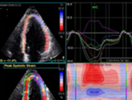
2D strain allows spatial and temporal tracking of longitudinal, circumferential and radial myocardial deformation. Its ability to provide angle-independent measures of LV strain is advantageous. Studies in patients with HCM have demonstrated a reduction in longitudinal strain with basal to apical gradient, an increase in circumferential strain, normal systolic twist or torsion, and reduction in untwisting in diastole. Another study showed significantly decreased longitudinal, circumferential, and radial strains in patients with HCM, compared to a healthy control group (23.3±17% vs. 27.2±14.9%, 16.8±7.1% vs. 19.65.2%, 25.2±13.9% vs. 36.8±17.2%, respectively. Significant reductions in strain in the septal segments (particularly the mid-septal segment), correlate with the septal/posterior wall ratio. (10) Longitudinal deformation abnormalities are often focal or subsegmental and may be underestimated if careful spatial mapping is not used. Because of its intrinsic ability to provide angle-independent strain data, 2D strain holds a unique advantage over tissue Doppler-derived strain, which is particularly important for myocardial deformation analysis in the apical LV segments.
9 - Three-dimensional echocardiography
3D-echocardiography has provided insights into the mechanics of SAM and deformational geometry of the LV outflow tract. This method facilitates the assessment of the LVOT area after intervention for septal reduction, surgical myectomy, volumetric estimates of left atrial mechanical function, and accurate estimation of LV ejection fraction, as well as LV mass in hypertrophied hearts (comparing favorably with CMR imaging).
10 - Transoesophageal echocardiography
Transoesophageal echocardiography should be considered in patients with poor transthoracic echo windows as an alternative or complementary investigation to CMR. It is particularly useful in patients with LVOTO if the mechanism is unclear when assessing the mitral valve apparatus before a septal reduction procedure and when severe mitral regurgitation caused by intrinsic valve abnormalities is suspected. In patients undergoing septal myectomy, perioperative TEE should be used to guide the surgical strategy and to detect surgical complications – ventricular septal defect and aortic regurgitation (AR)) and residual LVOTO. Following myectomy, intraoperative TEE studies can confirm the adequacy of myectomy and quantitate residual gradients, severity of mitral and aortic regurgitation, ventricular function, and development of a ventricular septal defect. (8)
11 - Contrast echocardiography
Contrast echocardiography currently is used to enhance endocardial definition, Doppler signals, and to evaluate myocardial perfusion. Myocardial contrast echocardiography can help anticipate the location and delineate the extent of septal infarct (selective injection of contrast into septal perforators) before alcohol septal ablation.
12 - Intraventricular dyssynchrony and prognosis
Interventricular and intraventricular delays are commonly present in patients with HCM despite the absence of conduction abnormalities on the electrocardiogram and appear to correlate to the degree of septal LVH and the presence of LV outflow obstruction. The prevalence and degree of LV dyssynchrony was significantly greater in patients with HCM compared with hypertensive LVH and age-matched control patients. Another study of 123 patients with HCM found that an intraventricular delay of >45 ms predicted an increased risk for ventricular tachyarrhythmias and sudden cardiac death at five-year follow-up (85.5% sensitivity; 90.4% specificity; positive predictive value: 66.9%; negative predictive value: 96.7%; test accuracy: 88.8%). (11,12)
13 - Echocardiographic predictors of unfavourable outcome
Several echocardiographic parameters given below are also important for prediction of unfavourable outcomes in HCM patients. (13)
- Sudden death: maximal wall thickness >30mm
- HCM related death: LVOT gradient at rest >30mmHg, EF <50%
- All-cause mortality: left atrial diameter >48mm
- Heart failure development: LA volume of >27ml/m2 at baseline during follow-up
- Intraventricular dyssynchrony: >45
14 - Differentiating HCM from athlete’s heart
Differential diagnosis between HCM and physiologic non-pathologic left ventricular (LV) hypertrophy associated with physical training (commonly referred to as 'athlete’s heart') has important implications because identification of cardiovascular disease associated with sudden death may be the basis for disqualification from competition to minimise risk. Conversely, improper overdiagnosis of cardiac disease may lead to unnecessary withdrawal from athletics. Echocardiography is one of the most important methods for differentiation between pathologic and physiologic left ventricular hypertrophy. (14)
15 - Differentiation between physiologic LHV and HCMP
Criteria favoring HCMP:
- Systolic mitral annual velocity <9m/sec
- Diastolic dysfunction
- Lack of LVH regression after cessation of exercise
- Lack of LV dilatation
- Provocable LVOT gradient
- Attenuated longitudinal, circumferential and radial strains (2D strain)
- Intraventricular dyssynchrony
Common diagnostic challenges include the following:
- Presentation in the late phase of the disease with a dilated and/orhypokinetic left ventricle and LV wall thinning
- Physiological hypertrophy caused by intense athletic training
- Patients with coexistent pathologies
- Isolated basal septal hypertrophy in elderly people
Conclusions
Conventional echocardiography (TTE) is the most readily accessible and informative tool in making the diagnosis of HCM. It is also the first noninvasive imaging method for risk stratification, treatment selection and follow-up of patients. Two-dimensional strain is a simple, rapid, and reproducible method to early detection of abnormalities in patients with HCM who have apparently normal left ventricular systolic function.

Figure 1. Parasternal long-axis view showing severe asymmetric septal hypertrophy.

Figure 2. M-mode across the mitral leaflets showing the presence of systolic anterior motion of the anterior mitral leaflet (SAM).

Figure 3. Late-peaking dynamic left ventricular outflow tract obstruction after the Valsalva maneuver.

Figure 4.Two-dimensional strain (quad format) images showing paradoxical apical longitudinal strain and corresponding abnormalities in the color M-mode of the parametric strain map. Loss of base-apex (strain) gradient is present.


 Our mission: To reduce the burden of cardiovascular disease.