In order to bring you the best possible user experience, this site uses Javascript. If you are seeing this message, it is likely that the Javascript option in your browser is disabled. For optimal viewing of this site, please ensure that Javascript is enabled for your browser.
Did you know that your browser is out of date? To get the best experience using our website we recommend that you upgrade to a newer version. Learn more.

Tricuspid regurgitation – Part 1: evaluation and risk stratification

Tricuspid regurgitation (TR) is a common finding in the general population with increasing prevalence in elderly patients, leading to impaired functional capacity, peripheral oedema, ascites, and congestive right heart failure with renal and hepatic impairment. Significant TR is associated with a higher cardiovascular mortality, even when adjusted for comorbidities. With better understanding of the tricuspid valve and the currently available transcatheter treatment options, the identification and screening of patients with relevant TR and referral to specialised centres is of increasing importance. This article describes the management of patients with relevant TR in terms of risk stratification (treatment options are addressed in a complementary article).

Valvular Heart Disease

Take-home messages


Introduction

Tricuspid regurgitation (TR) is a common finding during routine echocardiography [1]. Mild TR is detected in 75% of adults, while relevant (≥moderate) TR is estimated to affect 4% of the population over 75 years of age, which is comparable to the prevalence of aortic stenosis and mitral regurgitation [1-3]. For yet unknown reasons and in contrast to other valvular diseases, women are up to four times more frequently affected than men and the female sex is an independent predictor of the severity and progression of the disease [4-6]. This may be due to a higher prevalence of atrial fibrillation and heart failure with preserved ejection fraction (HFpEF) [7]. Besides age, sex and atrial fibrillation, pre- and postcapillary pulmonary hypertension are predictors of disease severity and progress [8].

TR manifests with impaired functional capacity and dyspnoea on exertion, as well as peripheral oedema, ascites, gastrointestinal symptoms and right heart failure, and in the advanced stages, consecutive renal and liver dysfunction [9]. The first therapeutic measure consists of the initiation of diuretic treatment.

Because of the longstanding and misleading belief that the treatment of left-sided heart disease improves TR in the majority of patients and that TR does not contribute to clinical symptoms and long-term outcomes, research on this topic has been scarce in the past [9,10]. In recent years, evidence has accumulated that significant TR is associated with an increased mortality of 40-70% after 1-4 years, even when adjusted for comorbidities [11-14]. These mortality rates are comparable to aortic stenosis [15] and results have been replicated in different study cohorts, applying not only to patients with isolated TR but also to patients with heart failure with reduced ejection fraction (HFrEF) [16].

Despite increasing evidence, TR remains a rarely assessed and treated valve disease and patients are often referred too late, when irreversible right heart failure is present and subsequent treatment is associated with increased operative risk.

Anatomical and echocardiographic considerations

The tricuspid valve (TV) is the largest and most anteriorly located heart valve separating the right atrium from the right ventricle with a complex and variable anatomy. Imaging is key in diagnosis and procedural planning. Only 54% of patients present the typical three-leaflet anatomy and around 1/3 have a  four-leaflet anatomy. Separation of the posterior leaflet is the most common variation, which increases the complexity of transcatheter treatment [17]. Furthermore, the commonly used echocardiographic grading for valvular regurgitation “mild-moderate-severe” does not adequately take into account the late stages of the disease and TR reduction after transcatheter treatment. In order to better address TR severity a new grading system has been proposed, adding two additional categories “massive” and “torrential” [18].


Figure 1. Echocardiographic evaluation and grading of tricuspid regurgitation (TR).

343_Dernetski_Figure 1_NEW.JPG

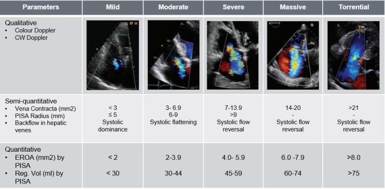
EROA: effective regurgitant orifice area; PISA: proximal isovelocity surface area


Classification and aetiology of TR

TR is classified according to its underlying pathomechanism into primary and secondary TR (STR). Primary TR (PTR) is rare, presenting in fewer than 10% of cases and is associated with abnormal leaflet morphology. Usually, PTR is observed in congenital or rheumatic heart disease or can be the consequence of infective endocarditis [19]. Infrequently repeated myocardial biopsies after (orthotropic) heart transplant can lead to PTR [20].

STR is the more prevalent pathomechanism and is due to changes in the atrial and/or ventricular geometry, leading to tethering of the leaflets or dilatation of the annulus [21] and is usually caused by left heart disease or pulmonary hypertension [22]. Atrial STR is newly recognised as a separate aetiology, due to long-standing atrial fibrillation (and age) with atrial remodelling [21]. With more cardiac electronic devices being implanted, cardiac implantable electronic devices (CIED)-related TR has increased and represent a distinct entity necessitating interdisciplinary approach and management [23,24].


Figure 2. Aetiological classification of TR.

343_Dernetski_Figure 2_NEW.JPG

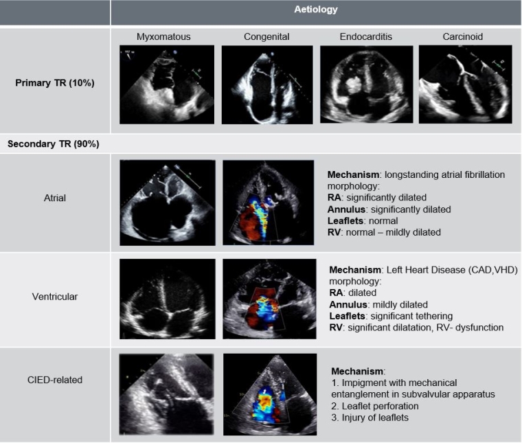
CAD: coronary artery disease; CIED: cardiac implantable electronic devices; RA: right atrium; RV: right ventricle; TR: tricuspid regurgitation; VHD: valvular heart disease


Risk stratification/ Guideline recommendations

With an ageing population and more minimally invasive treatment options available, the need to predict mortality, in order to adequately select patients who will benefit from an intervention or who are eligible for surgery, is crucial. Since neither the Society of Thoracic Surgeons (STS) score nor the European System for Cardiac Operative Risk Evaluation (EuroSCORE) II were designed to predict the risk of isolated TV interventions, a new dedicated score has been proposed for isolated tricuspid surgery [25,26] based on clinical predictors (Table 1) [25].


Figure 3. TRI-Score: prediction of in- hospital mortality after isolated TV-surgery.

343_Dernetski_Figure 3exTable1.jpg

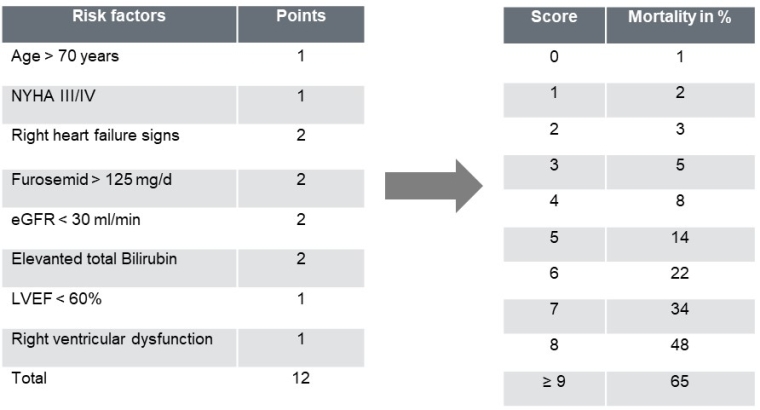
eGFR: estimated glomerular filtration rate; LVEF: left ventricular ejection fraction; NYHA: New York Heart Association


There is no consensus about the best strategy and patient population for isolated TV interventions. Current European guidelines recommend TV surgery for severe primary/secondary TR when undergoing left heart surgery (Class Ia indication). Isolated TV surgery is indicated for severe, symptomatic TR (Class Ia indication). Patients with severe, asymptomatic (or oligosymptomatic) TR without RV dysfunction or RV dilatation also have a Class IIa recommendation [31].

Conclusion

Isolated tricuspid valve surgery is still considered high risk with in-hospital mortality rates ~10% and presenting high mortality during follow-up (major adverse events occur in about ~30% of patients with advanced disease) [27-30]. These results correlate primarily with the severity of TR rather than its aetiology, leading to the conclusion that early referral is essential in order to improve outcomes.


Figure 4. Comprehensive approach to evaluate patients with TR with multimodal cardiac imaging and clinical assessment.

343_Dernetski_Figure 4_NEW.JPG

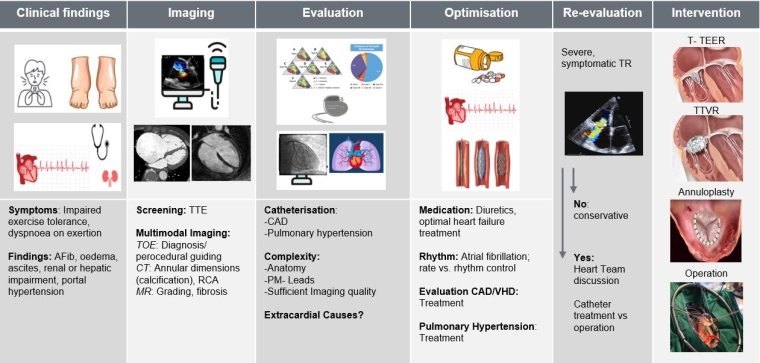
CAD: coronary artery disease; CT: computed tomography; MR: mitral regurgitation; RCA: right coronary artery; TOE: transoesophageal echocardiography; TR: tricuspid regurgitation; TTE: transthoracic echocardiography; T-TEER: tricuspid transcatheter edge-to-edge repair; TTVR: transcatheter tricuspid valve replacement; VHD: valvular heart disease


Impact on Practice

Severe, symptomatic tricuspid regurgitation has an important impact on clinical outcomes and needs to be addressed early in order to improve symptoms and quality of life, therefore early referral to a specialised centre is of major importance.

References


  1. Topilsky Y, Maltais S, Medina Inojosa J, Oguz D, Michelena H, Maalouf J, Mahoney DW, Enriquez-Sarano M. Burden of Tricuspid Regurgitation in Patients Diagnosed in the Community Setting. JACC Cardiovasc Imaging. 2019 Mar;12(3):433-442.
  2. Nkomo VT, Gardin JM, Skelton TN, Gottdiener JS, Scott CG, Enriquez-Sarano M. Burden of valvular heart diseases: a population-based study. Lancet. 2006 Sep 16;368(9540):1005-11.
  3. Cahill TJ, Prothero A, Wilson J, Kennedy A, Brubert J, Masters M, Newton JD, Dawkins S, Enriquez-Sarano M, Prendergast BD, Myerson SG. Community prevalence, mechanisms and outcome of mitral or tricuspid regurgitation. Heart. 2021 May 26;107(12):1003-1009.
  4. Singh JP, Evans JC, Levy D, Larson MG, Freed LA, Fuller DL, Lehman B, Benjamin EJ. Prevalence and clinical determinants of mitral, tricuspid, and aortic regurgitation (the Framingham Heart Study). Am J Cardiol. 1999 Mar 15;83(6):897-902.
  5. Offen S, Playford D, Strange G, Stewart S, Celermajer DS. Adverse Prognostic Impact of Even Mild or Moderate Tricuspid Regurgitation: Insights from the National Echocardiography Database of Australia. J Am Soc Echocardiogr. 2022 Aug;35(8):810-817..
  6. Beale AL, Nanayakkara S, Segan L, Mariani JA, Maeder MT, van Empel V, Vizi D, Evans S, Lam CSP, Kaye DM. Sex Differences in Heart Failure With Preserved Ejection Fraction Pathophysiology: A Detailed Invasive Hemodynamic and Echocardiographic Analysis. JACC Heart Fail. 2019 Mar;7(3):239-249.
  7. Mutlak D, Khalil J, Lessick J, Kehat I, Agmon Y, Aronson D. Risk Factors for the Development of Functional Tricuspid Regurgitation and Their Population-Attributable Fractions. JACC Cardiovasc Imaging. 2020 Aug;13(8):1643-1651.
  8. Arsalan M, Walther T, Smith RL 2nd, Grayburn PA. Tricuspid regurgitation diagnosis and treatment. Eur Heart J. 2017 Mar 1;38(9):634-638.
  9. Leurent G, Collet JP. The Tricuspid Valve: No Longer the Forgotten Valve! JACC Cardiovasc Interv. 2019 Dec 23;12(24):2496-2498.
  10. Reineke DC, Roost E, Schoenhoff F, Pasic M, Kadner A, Englberger L, Carrel TP. Transcatheter Treatment of Tricuspid Valve Disease: An Unmet Need? The Surgical Point of View. Front Cardiovasc Med. 2018 Jul 20;5:98.
  11. Nath J, Foster E, Heidenreich PA. Impact of tricuspid regurgitation on long-term survival. J Am Coll Cardiol. 2004 Feb 4;43(3):405-9.
  12. Neuhold S, Huelsmann M, Pernicka E, Graf A, Bonderman D, Adlbrecht C, Binder T, Maurer G, Pacher R, Mascherbauer J. Impact of tricuspid regurgitation on survival in patients with chronic heart failure: unexpected findings of a long-term observational study. Eur Heart J. 2013 Mar;34(11):844-52.
  13. Samim D, Praz F, Cochard B, Brugger N, Ruberti A, Bartkowiak J, Corpataux N, Reineke D, Pilgrim T, Windecker S, Wenaweser PM, Wild MG. Natural history and mid-term prognosis of severe tricuspid regurgitation: A cohort study. Front Cardiovasc Med. 2023 Jan 9;9:1026230.
  14. Kadri AN, Menon V, Sammour YM, Gajulapalli RD, Meenakshisundaram C, Nusairat L, Mohananey D, Hernandez AV, Navia J, Krishnaswamy A, Griffin B, Rodriguez L, Harb SC, Kapadia S. Outcomes of patients with severe tricuspid regurgitation and congestive heart failure. Heart. 2019 Dec;105(23):1813-1817.
  15. Leon MB, Smith CR, Mack M, Miller DC, Moses JW, Svensson LG, Tuzcu EM, Webb JG, Fontana GP, Makkar RR, Brown DL, Block PC, Guyton RA, Pichard AD, Bavaria JE, Herrmann HC, Douglas PS, Petersen JL, Akin JJ, Anderson WN, Wang D, Pocock S; PARTNER Trial Investigators. Transcatheter aortic-valve implantation for aortic stenosis in patients who cannot undergo surgery. N Engl J Med. 2010 Oct 21;363(17):1597-607.
  16. Benfari G, Antoine C, Miller WL, Thapa P, Topilsky Y, Rossi A, Michelena HI, Pislaru S, Enriquez-Sarano M. Excess Mortality Associated With Functional Tricuspid Regurgitation Complicating Heart Failure With Reduced Ejection Fraction. Circulation. 2019 Jul 16;140(3):196-206.
  17. Hahn RT, Weckbach LT, Noack T, Hamid N, Kitamura M, Bae R, Lurz P, Kodali SK, Sorajja P, Hausleiter J, Nabauer M. Proposal for a Standard Echocardiographic Tricuspid Valve Nomenclature. JACC Cardiovasc Imaging. 2021 Jul;14(7):1299-1305.
  18. Hahn RT, Zamorano JL. The need for a new tricuspid regurgitation grading scheme. Eur Heart J Cardiovasc Imaging. 2017 Dec 1;18(12):1342-1343.
  19. Condello F, Gitto M, Stefanini GG. Etiology, epidemiology, pathophysiology and management of tricuspid regurgitation: an overview. Rev Cardiovasc Med. 2021 Dec 22;22(4):1115-1142.
  20. Fiorelli AI, Coelho GH, Aiello VD, Benvenuti LA, Palazzo JF, Santos Júnior VP, Canizares B, Dias RR, Stolf NA. Tricuspid valve injury after heart transplantation due to endomyocardial biopsy: an analysis of 3550 biopsies. Transplant Proc. 2012 Oct;44(8):2479-82.
  21. Hahn RT, Lawlor MK, Davidson CJ, Badhwar V, Sannino A, Spitzer E, Lurz P, Lindman BR, Topilsky Y, Baron SJ, Chadderdon S, Khalique OK, Tang GHL, Taramasso M, Grayburn PA, Badano L, Leipsic J, Lindenfeld J, Windecker S, Vemulapalli S, Redfors B, Alu MC, Cohen DJ, Rodés-Cabau J, Ailawadi G, Mack M, Ben-Yehuda O, Leon MB, Hausleiter J; TVARC Steering Committee. Tricuspid Valve Academic Research Consortium Definitions for Tricuspid Regurgitation and Trial Endpoints. J Am Coll Cardiol. 2023 Oct 24;82(17):1711-1735.
  22. Badano LP, Muraru D, Enriquez-Sarano M. Assessment of functional tricuspid regurgitation. Eur Heart J. 2013 Jul;34(25):1875-85.
  23. Hahn RT. Tricuspid Regurgitation. N Engl J Med. 2023 May 18;388(20):1876-1891.
  24. Praz F, Muraru D, Kreidel F, Lurz P, Hahn RT, Delgado V, Senni M, von Bardeleben RS, Nickenig G, Hausleiter J, Mangieri A, Zamorano JL, Prendergast BD, Maisano F. Transcatheter treatment for tricuspid valve disease. EuroIntervention. 2021 Nov 19;17(10):791-808.
  25. Dreyfus J, Audureau E, Bohbot Y, Coisne A, Lavie-Badie Y, Bouchery M, Flagiello M, Bazire B, Eggenspieler F, Viau F, Riant E, Mbaki Y, Eyharts D, Senage T, Modine T, Nicol M, Doguet F, Nguyen V, Le Tourneau T, Tribouilloy C, Donal E, Tomasi J, Habib G, Selton-Suty C, Raffoul R, Iung B, Obadia JF, Messika-Zeitoun D. TRI-SCORE: a new risk score for in-hospital mortality prediction after isolated tricuspid valve surgery. Eur Heart J. 2022 Feb 12;43(7):654-662.
  26. Selton-Suty C, Radu C, Lim P, Raffoul R, Iung B, Obadia JF, Audureau E, Messika-Zeitoun D. Predictive value of the TRI-SCORE for in-hospital mortality after redo isolated tricuspid valve surgery. Heart. 2023 May 26;109(12):951-958.
  27. Dreyfus J, Ghalem N, Garbarz E, Cimadevilla C, Nataf P, Vahanian A, Caranhac G, Messika-Zeitoun D. Timing of Referral of Patients With Severe Isolated Tricuspid Valve Regurgitation to Surgeons (from a French Nationwide Database). Am J Cardiol. 2018 Jul 15;122(2):323-326.
  28. Scotti A, Sturla M, Granada JF, Kodali SK, Coisne A, Mangieri A, Godino C, Ho E, Goldberg Y, Chau M, Jorde UP, Garcia MJ, Maisano F, Bapat VN, Ailawadi G, Latib A. Outcomes of isolated tricuspid valve replacement: a systematic review and meta-analysis of 5,316 patients from 35 studies. EuroIntervention. 2022 Nov 18;18(10):840-851.
  29. Kilic A, Saha-Chaudhuri P, Rankin JS, Conte JV. Trends and outcomes of tricuspid valve surgery in North America: an analysis of more than 50,000 patients from the Society of Thoracic Surgeons database. Ann Thorac Surg. 2013 Nov;96(5):1546-52; discussion 1552.
  30. Axtell AL, Bhambhani V, Moonsamy P, Healy EW, Picard MH, Sundt TM 3rd, Wasfy JH. Surgery Does Not Improve Survival in Patients With Isolated Severe Tricuspid Regurgitation. J Am Coll Cardiol. 2019 Aug 13;74(6):715-725.
  31. Vahanian A, Beyersdorf F, Praz F, Milojevic M, Baldus S, Bauersachs J, Capodanno D, Conradi L, De Bonis M, De Paulis R, Delgado V, Freemantle N, Gilard M, Haugaa KH, Jeppsson A, Jüni P, Pierard L, Prendergast BD, Sádaba JR, Tribouilloy C, Wojakowski W; ESC/EACTS Scientific Document Group. 2021 ESC/EACTS Guidelines for the management of valvular heart disease: Developed by the Task Force for the management of valvular heart disease of the European Society of Cardiology (ESC) and the European Association for Cardio-Thoracic Surgery (EACTS). Rev Esp Cardiol (Engl Ed). 2022 Jun;75(6):524.

Notes to editor


Authors:

Chrisoula Dernektsi1, MD; Tetsu Tanaka2, BS, MS PhD; Johanna Vogelhuber2, MD; Georg Nickenig2, MD, PhD; Fabien Praz1, MD

Affiliations:

  1. University Hospital Bern, Department of Cardiology, Inselspital, University of Bern, Bern, Switzerland;
  2. Heart Center Bonn, Department of Cardiology, University Hospital Bonn, Bonn, Germany


Address for correspondence:

Dr Chrisoula Dernektsi, Universitätsklinik für Kardiologie, Inselspital Bern, 3010 Bern, Switzerland

Phone: +41 31 632 50 00


Email: chrisoula.dernektsi@insel.ch


Author disclosures:

Chrisoula Dernektsi: No Conflict of Interest

Fabien Praz: Fabien Praz was compensated for travel expenses by Abbott Vascular, Edwards Lifesciences, Medira, Polares Medical und Siemens Healthineers

Georg Nickenig: Research funding from the Deutsche Forschungsgemeinschaft, the German Federal Ministry of Education and Research, the EU, Abbott, Edwards Lifesciences, Medtronic, and St Jude Medical. Honoraria for lectures or advisory boards from Abbott, Edwards Lifesciences, Medtronic, and St Jude Medical. 

Tetsu Tanaka: No Conflict of Interest

Johanna Vogelhuber: No Conflict of Interest


The content of this article reflects the personal opinion of the author/s and is not necessarily the official position of the European Society of Cardiology.